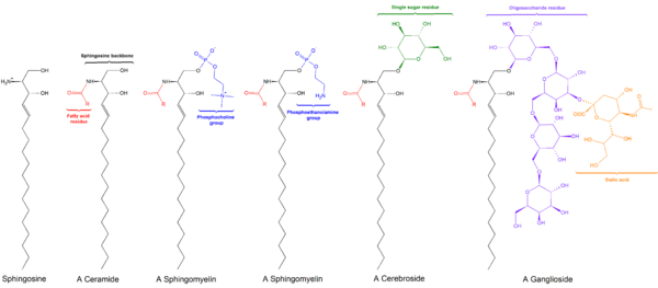
Ceramide


Ceramides are a family of waxy lipid molecules. A ceramide is composed of sphingosine and a fatty acid. Ceramides are found in high concentrations within the cell membrane of cells, since they are component lipids that make up sphingomyelin, one of the major lipids in the lipid bilayer. Contrary to previous assumptions that ceramides and other sphingolipids found in cell membrane were purely supporting structural elements, ceramide can participate in a variety of cellular signaling: examples include regulating differentiation, proliferation, and programmed cell death (PCD) of cells.
The word ceramide comes from the Latin cera (wax) and amide. Ceramide is a component of vernix caseosa, the waxy or cheese-like white substance found coating the skin of newborn human infants.
Pathways for ceramide synthesis
There are three major pathways of ceramide generation. The sphingomyelinase pathway uses an enzyme to break down sphingomyelin in the cell membrane and release ceramide. The de novo pathway creates ceramide from less complex molecules. Ceramide generation can also occur through breakdown of complex sphingolipids that are ultimately broken down into sphingosine, which is then reused by reacylation to form ceramide. This latter pathway is termed the Salvage pathway.
Sphingomyelin hydrolysis
Hydrolysis of sphingomyelin is catalyzed by the enzyme sphingomyelinase. Because sphingomyelin is one of the four common phospholipids found in the plasma membrane of cells, the implications of this method of generating ceramide is that the cellular membrane is the target of extracellular signals leading to programmed cell death. There has been research suggesting that when ionizing radiation causes apoptosis in some cells, the radiation leads to the activation of sphingomyelinase in the cell membrane and ultimately, to ceramide generation.[1]
De novo
De novo synthesis of ceramide begins with the condensation of palmitate and serine to form 3-keto-dihydrosphingosine. This reaction is catalyzed by the enzyme serine palmitoyl transferase and is the rate-limiting step of the pathway. In turn, 3-keto-dihydrosphingosine is reduced to dihydrosphingosine, which is then followed by acylation by the enzyme (dihydro)ceramide synthase to produce dihydroceramide. The final reaction to produce ceramide is catalyzed by dihydroceramide desaturase. De novo synthesis of ceramide occurs in the endoplasmic reticulum. Ceramide is subsequently transported to the Golgi apparatus by either vesicular trafficking or the ceramide transfer protein CERT. Once in the Golgi apparatus, ceramide can be further metabolized to other sphingolipids, such as sphingomyelin and the complex glycosphingolipids.[2]
Salvage pathway
Constitutive degradation of sphingolipids and glycosphingolipids takes place in the acidic subcellular compartments, the late endosomes and the lysosomes, with the end goal of producing sphingosine. In the case of glycosphingolipids, exohydrolases acting at acidic pH optima cause the stepwise release of monosaccharide units from the end of the oligosaccharide chains, leaving just the sphingosine portion of the molecule, which may then contribute to the generation of ceramides. Ceramide can be further hydrolyzed by acid ceramidase to form sphingosine and a free fatty acid, both of which are able to leave the lysosome, unlike ceramide. The long-chain sphingoid bases released from the lysosome may then re-enter pathways for synthesis of ceramide and/or sphingosine-1-phosphate. The salvage pathway re-utilizes long-chain sphingoid bases to form ceramide through the action of ceramide synthase. Thus, ceramide synthase family members probably trap free sphingosine released from the lysosome at the surface of the endoplasmic reticulum or in endoplasmic reticulum-associated membranes. It should also be noted that the salvage pathway has been estimated to contribute from 50% to 90% of sphingolipid biosynthesis[3]
Physiological roles of ceramide
As a bioactive lipid, ceramide has been implicated in a variety of physiological functions including apoptosis, cell growth arrest, differentiation, cell senescence, cell migration and adhesion.[2] Roles for ceramide and its downstream metabolites have also been suggested in a number of pathological states including cancer, neurodegeneration, diabetes, microbial pathogenesis, obesity, and inflammation.[4][5]
Apoptosis
One of the most studied roles of ceramide pertains to its function as a proapoptotic molecule. Apoptosis, or Type I programmed cell death, is essential for the maintenance of normal cellular homeostasis and is an important physiological response to many forms of cellular stress. Ceramide accumulation has been found following treatment of cells with a number of apoptotic agents including ionizing radiation,[1][6] UV light,[7] TNF-alpha,[8] and chemotherapeutic agents. This suggests a role for ceramide in the biological responses of all these agents. Because of its apoptosis-inducing effects in cancer cells, ceramide has been termed the “tumor suppressor lipid” . Several studies have attempted to define further the specific role of ceramide in the events of cell death and some evidence suggests ceramide functions upstream of the mitochondria in inducing apoptosis. However, owing to the conflicting and variable nature of studies into the role of ceramide in apoptosis, the mechanism by which this lipid regulates apoptosis remains elusive.[9]
Skin
Ceramide is the main component of the stratum corneum of the epidermis layer of human skin.[10][11] Together with cholesterol and saturated fatty acids, ceramide creates a water-impermeable, protective organ to prevent excessive water loss due to evaporation as well as a barrier against the entry of microorganisms.[11] With aging there is a decline in ceramide and cholesterol in the stratum corneum of humans.[12]
Hormonal
Increased ceramide synthesis leads to both leptin resistance and insulin resistance by increasing SOCS-3 expression.[13] Elevated level of ceramide results in the inhibition of insulin signal transduction pathway and the serine phosphorylation of JNK, leading to insulin resistance.[14]
Substances known to induce ceramide generation
- Anandamide
- Ceramidase Inhibitors
- Chemotherapeutic agents
- Fas ligand
- Endotoxin
- homocysteine[15]
- heat
- gamma interferon
- ionizing radiation[1][16]
- matrix metalloproteinases[15]
- reactive oxygen species[15]
- Tetrahydrocannabinol and other Cannabinoids[17]
- TNF-alpha[15]
- 1,25 dihydroxy vitamin D
It is interesting to note that the substances that can cause ceramide to be generated tend to be stress signals that can cause the cells to go into programmed cell death. Ceramide thus acts as an intermediary signal that connects the external signal to the internal metabolism of the cells.
Mechanism by which ceramide signaling occurs
Currently, the means by which ceramide acts as a signaling molecule are not clear.
One hypothesis is that ceramide generated in the plasma membrane enhances membrane rigidity and stabilizes smaller lipid platforms known as lipid rafts, allowing them to serve as platforms for signalling molecules. Moreover, as rafts on one leaflet of the membrane can induce localized changes in the other leaflet of the bilayer, they can potentially serve as the link between signals from outside the cell to signals to be generated within the cell.
Ceramide has also been shown to form organized large channels traversing the mitochondrial outer membrane. This leads to the egress of proteins from the intermembrane space.[18][19][20]
Uses
Ceramides may be found as ingredients of some topical skin medications used to complement treatment for skin conditions such as eczema.[21] They are also used in cosmetic products such as some soaps, shampoos, skin creams, and sunscreens.[22] Additionally, ceramides are being explored as a potential therapeutic in cancer. [23]
References
- 1 2 3 Haimovitz-Friedman A, Kan CC, Ehleiter D, et al. (1994). "Ionizing radiation acts on cellular membranes to generate ceramide and initiate apoptosis". J. Exp. Med. 180 (2): 525–35. doi:10.1084/jem.180.2.525. PMC 2191598
 . PMID 8046331.
 - 1 2 Hannun, Y.A.; Obeid, L.M. (2008). "Principles of bioactive lipid signalling: lessons from sphingolipids". Nature Reviews Molecular Cell Biology. 9 (2): 139–150. doi:10.1038/nrm2329. PMID 18216770.
- ↑ Kitatani K, Idkowiak-Baldys J, Hannun YA (2008). "The sphingolipid salvage pathway in ceramide metabolism and signaling". Cell Signaling. 30 (6): 1010–1018. doi:10.1016/j.cellsig.2007.12.006. PMC 2422835
 . PMID 18191382.
 - ↑ Zeidan, Y.H.; Hannun, Y.A. (2007). "Translational aspects of sphingolipid metabolism". Trends Mol. Med. 13 (8): 327–336. doi:10.1016/j.molmed.2007.06.002. PMID 17588815.
- ↑ Wu D, Ren Z, Pae M, Guo W, Cui X, Merrill AH, Meydani SN (2007). "Aging up-regulates expression of inflammatory mediators in mouse adipose tissue". The Journal of Immunology. 179 (7): 4829–39. doi:10.4049/jimmunol.179.7.4829. PMID 17878382.
- ↑ Dbaibo GS, Pushkareva MY, Rachid RA, Alter N, Smyth MJ, Obeid LM, Hannun YA (1998). "p53-dependent ceramide response to genotoxic stress". J. Clin. Invest. 102 (2): 329–339. doi:10.1172/JCI1180. PMC 508891
 . PMID 9664074.
 - ↑ Rotolo JA, Zhang J, Donepudi M, Lee H, Fuks Z, Kolesnick R (2005). "Caspase-dependent and -independent activation of acid sphingomyelinase signaling". J. Biol. Chem. 280 (28): 26425–34. doi:10.1074/jbc.M414569200. PMID 15849201.
- ↑ Dbaibo GS, El-Assaad W, Krikorian A, Liu B, Diab K, Idriss NZ, El-Sabban M, Driscoll TA, Perry DK, Hannun YA (2001). "Ceramide generation by two distinct pathways in tumor necrosis factor alpha-induced cell death". FEBS Letters. 503 (1): 7–12. doi:10.1016/S0014-5793(01)02625-4. PMID 11513845.
- ↑ Taha TA, Mullen TD, Obeid LM (2006). "A house divided: ceramide, sphingosine, and sphingosine-1-phosphate in programmed cell death". Biochim. Biophys. Acta. 1758 (12): 2027–36. doi:10.1016/j.bbamem.2006.10.018. PMC 1766198
 . PMID 17161984.
 - ↑ Hill JR, Wertz PW (2009). "Structures of the ceramides from porcine palatal stratum corneum". LIPIDS. 44 (3): 291–295. doi:10.1007/s11745-009-3283-9. PMID 19184160.
- 1 2 Garidel P, Fölting B, Schaller I, Kerth A (2010). "The microstructure of the stratum corneum lipid barrier: mid-infrared spectroscopic studies of hydrated ceramide:palmitic acid:cholesterol model systems". Biophysical Chemistry. 150 (1-3): 144–156. doi:10.1016/j.bpc.2010.03.008. PMID 20457485.
- ↑ Popa I, Abdul-Malak N, Portoukalian J (2010). "The weak rate of sphingolipid biosynthesis shown by basal keratinocytes isolated from aged vs. young donors is fully rejuvenated after treatment with peptides of a potato hydrolysate". International Journal of Cosmetic Science. 32 (3): 225–232. doi:10.1111/j.1468-2494.2009.00571.x. PMID 20384897.
- ↑ Yang G, Badeanlou L, Bielawski J, Roberts AJ, Hannun YA, Samad F (2009). "Central role of ceramide biosynthesis in body weight regulation, energy metabolism, and the metabolic syndrome". American Journal of Physiology. 297 (1): E211–E224. doi:10.1152/ajpendo.91014.2008. PMC 2711669
 . PMID 19435851.
 - ↑ Febbraio, Mark (2014). "Role of interleukins in obesity:implications for metabolic disease". Trends in Endocrinology and Metabolism. 25 (6): 312–319.
- 1 2 3 4 Bismuth J, Lin P, Yao Q, Chen C (2008). "Ceramide: a common pathway for atherosclerosis?". Atherosclerosis (journal). 196 (2): 497–504. doi:10.1016/j.atherosclerosis.2007.09.018. PMC 2924671
 . PMID 17963772.
 - ↑ Hallahan DE (1996). "Radiation-mediated gene expression in the pathogenesis of the clinical radiation response". Sem. Radiat. Oncol. 6 (4): 250–267. doi:10.1016/S1053-4296(96)80021-X. PMID 10717183.
- ↑ Velasco, G; Galve-Roperh, I; Sánchez, C; Blázquez, C; Haro, A; Guzmán, M (2005). "Cannabinoids and ceramide: Two lipids acting hand-by-hand". Life Sciences. 77 (14): 1723–31. doi:10.1016/j.lfs.2005.05.015. PMID 15958274.
- ↑ Siskind LJ, Kolesnick RN, Colombini M (2002). "Ceramide Channels Increase the Permeability of the Mitochondrial Outer Membrane to Small Proteins". J. Biol. Chem. 277 (30): 26796–803. doi:10.1074/jbc.M200754200. PMC 2246046
 . PMID 12006562.
 - ↑ Stiban J, Fistere D, Colombini M (2006). "Dihydroceramide hinders ceramide channel formation: Implications on apoptosis". Apoptosis. 11 (5): 773–80. doi:10.1007/s10495-006-5882-8. PMID 16532372.
- ↑ Siskind LJ, Kolesnick RN, Colombini M (2006). "Ceramide forms channels in mitochondrial outer membranes at physiologically relevant concentrations". Mitochondrion. 6 (3): 118–25. doi:10.1016/j.mito.2006.03.002. PMC 2246045
 . PMID 16713754.
 - ↑ "Ceramides - Skin Lipids That Keep Skin Moisturized". Retrieved 29 January 2015.
- ↑ "Safety Assessment of Ceramides as Used in Cosmetics" (PDF). Cosmetic Ingredient Review. May 16, 2014.
- ↑ Huang, WC; Chen, CL; Lin, YS; Lin, CF (2011). "Apoptotic Sphingolipid Ceramide in Cancer Therapy". Journal of Lipids (2011): 1–15.
External links
- Ceramides at the US National Library of Medicine Medical Subject Headings (MeSH)