Tissue microarray


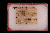
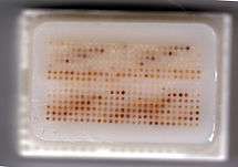
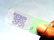
Tissue microarrays (also TMAs) consist of paraffin blocks in which up to 1000[1] separate tissue cores are assembled in array fashion to allow multiplex histological analysis.
History
The major limitations in molecular clinical analysis of tissues include the cumbersome nature of procedures, limited availability of diagnostic reagents and limited patient sample size. The technique of tissue microarray was developed to address these issues.
Multi-tissue blocks were first introduced by H. Battifora in 1986 with his so-called “multitumor (sausage) tissue block" and modified in 1990 with its improvement, "the checkerboard tissue block" . In 1998, J. Kononen and collaborators developed the current technique, which uses a novel sampling approach to produce tissues of regular size and shape that can be more densely and precisely arrayed.
Procedure
In the tissue microarray technique, a hollow needle is used to remove tissue cores as small as 0.6 mm in diameter from regions of interest in paraffin-embedded tissues such as clinical biopsies or tumor samples. These tissue cores are then inserted in a recipient paraffin block in a precisely spaced, array pattern. Sections from this block are cut using a microtome, mounted on a microscope slide and then analyzed by any method of standard histological analysis. Each microarray block can be cut into 100 – 500 sections, which can be subjected to independent tests. Tests commonly employed in tissue microarray include immunohistochemistry, and fluorescent in situ hybridization. Tissue microarrays are particularly useful in analysis of cancer samples.
One variation is a Frozen tissue array.
See also
References
- Battifora H: The multitumor (sausage) tissue block: novel method for immunohistochemical antibody testing. Lab Invest 1986, 55:244-248.
- Battifora H, Mehta P: The checkerboard tissue block. An improved multitissue control block. Lab Invest 1990, 63:722-724.
- Kononen J, Bubendorf L, Kallioniemi A, Barlund M, Schraml P, Leighton S, Torhorst J, Mihatsch MJ, Sauter G, Kallioniemi OP: Tissue microarrays for high-throughput molecular profiling of tumor specimens. Nat Med 1998, 4:844-847.