Mandible
| Mandible | |
|---|---|
 The skull from the front, with mandible shown in purple at bottom. | |
| Details | |
| Precursor | 1st branchial arch[1] |
| Identifiers | |
| Latin | mandibula |
| MeSH | Mandible |
| FMA | 52748 |
The mandible,[2] lower jaw or jawbone (from Latin mandibula, "jawbone") is the largest, strongest and lowest bone in the face.[3] It forms the lower jaw and holds the lower teeth in place. In the midline on the anterior surface of the mandible is a faint ridge, an indication of the mandibular symphysis, where the bone is formed by the fusion of right and left processes during mandibular development. Like other symphyses in the body, this is a midline articulation where the bones are joined by fibrocartilage, but this articulation fuses together in early childhood.[4]
Structure


The mandible consists of:
- a curved, horizontal portion, the body or base. (See body of mandible).
- two perpendicular parts, the rami, or ramus for each one, unite with the ends of the body nearly at right angles. (See ramus mandibulae). The angle formed at this junction is called the angle of the mandible or the gonial angle.
- Alveolar process, the tooth bearing area of the mandible (upper part of the body of the mandible)
- Condyle, superior (upper) and posterior projection from the ramus, which makes the temporomandibular joint with the temporal bone
- Coronoid process, superior and anterior projection from the ramus. This provides attachment to the temporalis muscle
The mandible articulates with the two temporal bones at the temporomandibular joints.
Foramina
- Mandibular foramen, paired, in the inner (medial) aspect of the mandible, superior to the mandibular angle in the middle of the ramus.
- Mental foramen, paired, lateral to the mental protuberance (chin) on the body of mandible, usually inferior to the apices of the mandibular first and second premolars. As mandibular growth proceeds in young children, the mental foramen alters in direction of its opening from anterior to posterosuperior. The mental foramen allows the entrance of the mental nerve and blood vessels into the mandibular canal.[4]
Nerves
Inferior alveolar nerve, branch of the mandibular division of Trigeminal (V) nerve, enters the mandibular foramen and runs forward in the mandibular canal, supplying sensation to the teeth. At the mental foramen the nerve divides into two terminal branches: incisive and mental nerves. The incisive nerve runs forward in the mandible and supplies the anterior teeth. The mental nerve exits the mental foramen and supplies sensation to the lower lip.
Rarely, a bifid inferior alveolar nerve may be present, in which case a second mandibular foramen, more inferiorly placed, exists and can be detected by noting a doubled mandibular canal on a radiograph.[4]
Development
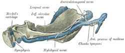
The ossification of the mandible refers to the Human mandible laying down new bone material in the fibrous membrane covering the outer surfaces of Meckel's cartilages.
These cartilages form the cartilaginous bar of the mandibular arch, and are two in number, a right and a left. Their proximal or cranial ends are connected with the ear capsules, and their distal extremities are joined to one another at the symphysis by mesodermal tissue. They run forward immediately below the condyles and then, bending downward, lie in a groove near the lower border of the bone; in front of the canine tooth they incline upward to the symphysis. From the proximal end of each cartilage the malleus and incus, two of the bones of the middle ear, are developed; the next succeeding portion, as far as the lingula, is replaced by fibrous tissue, which persists to form the sphenomandibular ligament.
Between the lingula and the canine tooth the cartilage disappears, while the portion of it below and behind the incisor teeth becomes ossified and incorporated with this part of the mandible.
Ossification takes place in the membrane covering the outer surface of the ventral end of Meckel's cartilage (Figs. 178 to 181), and each half of the bone is formed from a single center which appears, near the mental foramen, about the sixth week of fetal life.
By the tenth week the portion of Meckel's cartilage which lies below and behind the incisor teeth is surrounded and invaded by the membrane bone. Somewhat later, accessory nuclei of cartilage make their appearance:
- a wedge-shaped nucleus in the condyloid process and extending downward through the ramus;
- a small strip along the anterior border of the coronoid process;
- smaller nuclei in the front part of both alveolar walls and along the front of the lower border of the bone.
These accessory nuclei possess no separate ossific centers, but are invaded by the surrounding membrane bone and undergo absorption. The inner alveolar border, usually described as arising from a separate ossific center (splenial center), is formed in the human mandible by an ingrowth from the main mass of the bone.
At birth the bone consists of two parts, united by a fibrous symphysis, in which ossification takes place during the first year.
 Figure 3: Mandible of human embryo 24 mm. long. Outer aspect.
 Figure 4: Mandible of human embryo 24 mm. long. Inner aspect.
 Figure 5: Mandible of human embryo 95 mm. long. Outer aspect. Nuclei of cartilage stippled.
 Figure 5: Mandible of human embryo 95 mm. long. Inner aspect. Nuclei of cartilage stippled.
Changes by age
When remains of humans are found, the mandible is one of the common findings, sometimes the only bone found. Skilled experts can estimate the age of the human upon death because the mandible changes over a person's life, as described in this article.
At birth, the body of the bone is a mere shell, containing the sockets of the two incisor, the canine, and the two deciduous molar teeth, imperfectly partitioned off from one another. The mandibular canal is of large size and runs near the lower border of the bone; the mental foramen opens beneath the socket of the first deciduous molar tooth. The angle is obtuse (175°), and the condyloid portion is nearly in line with the body. The coronoid process is of comparatively large size, and projects above the level of the condyle.
After birth, the two segments of the bone become joined at the symphysis, from below upward, in the first year; but a trace of separation may be visible in the beginning of the second year, near the alveolar margin. The body becomes elongated in its whole length, but more especially behind the mental foramen, to provide space for the three additional teeth developed in this part. The depth of the body increases owing to increased growth of the alveolar part, to afford room for the roots of the teeth, and by thickening of the subdental portion which enables the jaw to withstand the powerful action of the masticatory muscles; but, the alveolar portion is the deeper of the two, and, consequently, the chief part of the body lies above the oblique line. The mandibular canal, after the second dentition, is situated just above the level of the mylohyoid line; and the mental foramen occupies the position usual to it in the adult. The angle becomes less obtuse, owing to the separation of the jaws by the teeth; about the fourth year it is 140°.
In the adult, the alveolar and subdental portions of the body are usually of equal depth. The mental foramen opens midway between the upper and lower borders of the bone, and the mandibular canal runs nearly parallel with the mylohyoid line. The ramus is almost vertical in direction, the angle measuring from 110° to 120°, also the adult condyle is higher than the coronoid process and the sigmoid notch becomes deeper.
In old age, the bone becomes greatly reduced in volume due to the loss of teeth and consequent resorption of the alveolar processes and interalveolar septa. Consequently, the chief part of the bone is below the oblique line. The mandibular canal, with the mental foramen opening from it, is closer to the alveolar border. The ramus is oblique in direction, the angle measures about 140°, and the neck of the condyle is more or less bent backward.
 Fig. 1: At birth.
 Fig. 2: In childhood.
 Fig. 3: In the adult.
 Fig. 4: In old age. Side view of the mandible at different periods of life.
Variation
Males generally have squarer, stronger, and larger mandibles than females. The mental protuberance is more pronounced in males but can be visualized and palpated in females.
Clinical relevance
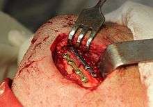
One fifth of facial injuries involve mandibular fracture.[5] Mandibular fractures are often accompanied by a 'twin fracture' on the contralateral (opposite) side. There is no universally accepted treatment protocol, as there is no consensus on the choice of techniques in a particular anatomical shape of mandibular fracture clinic. A common treatment involves attachment of metal plates to the fracture to assist in healing.[6]
Mandibular fractures

- Motor vehicle accident (MVA) – 40%
- Assault – 40%
- Fall – 10%
- Sport – 5%
- Other – 5%
Location of mandibular fractures
- Condyle – 30%
- Angle – 25%
- Body – 25%
- Symphysis – 15%
- Ramus – 3%
- Coronoid process – 2%[7]
The mandible may be dislocated anteriorly (to the front) and inferiorly (downwards) but very rarely posteriorly (backwards).
The mandibular alveolar process can become resorbed when completely edentulous in the mandibular arch (occasionally noted also in partially edentulous cases). This resorption can occur to such an extent that the mental foramen is virtually on the superior border of the mandible, instead of opening on the anterior surface, changing its relative position. However, the more inferior body of the mandible is not affected and remains thick and rounded. With age and tooth loss, the alveolar process is absorbed so that the mandibular canal becomes nearer the superior border. Sometimes with excessive alveolar process absorption, the mandibular canal disappears entirely and leaves the inferior alveolar nerve without its bony protection, although it is still covered by soft tissue.[4]
In other vertebrates
.jpg)
In lobe-finned fishes and the early fossil tetrapods, the bone homologous to the mandible of mammals is merely the largest of several bones in the lower jaw. In such animals, it is referred to as the dentary bone, and forms the body of the outer surface of the jaw. It is bordered below by a number of splenial bones, while the angle of the jaw is formed by a lower angular bone and a suprangular bone just above it. The inner surface of the jaw is lined by a prearticular bone, while the articular bone forms the articulation with the skull proper. Finally a set of three narrow coronoid bones lie above the prearticular bone. As the name implies, the majority of the teeth are attached to the dentary, but there are commonly also teeth on the coronoid bones, and sometimes on the prearticular as well.[8]
This complex primitive pattern has, however, been simplified to various degrees in the great majority of vertebrates, as bones have either fused or vanished entirely. In teleosts, only the dentary, articular, and angular bones remain, while in living amphibians, the dentary is accompanied only by the prearticular, and, in salamanders, one of the coronoids. The lower jaw of reptiles has only a single coronoid and splenial, but retains all the other primitive bones except the prearticular and the periosteum.[8]
While, in birds, these various bones have fused into a single structure, in mammals most of them have disappeared, leaving an enlarged dentary as the only remaining bone in the lower jaw - the mandible. As a result of this, the primitive jaw articulation, between the articular and quadrate bones, has been lost, and replaced with an entirely new articulation between the mandible and the temporal bone. An intermediate stage can be seen in some therapsids, in which both points of articulation are present. Aside from the dentary, only few other bones of the primitive lower jaw remain in mammals; the former articular and quadrate bones survive as the malleus and the incus of the middle ear.[8]
Finally, the cartilaginous fish, such as sharks, do not have any of the bones found in the lower jaw of other vertebrates. Instead, their lower jaw is composed of a cartilagenous structure homologous with the Meckel's cartilage of other groups. This also remains a significant element of the jaw in some primitive bony fish, such as sturgeons.[8]
Mandible in popular culture
- In the Book of Judges, Samson used a donkey's jawbone to kill a thousand Philistines.[9]
Additional images
 Lateral view
 Front
Front Mandible
 Gray181.png
 The surgical treatment of mandibular angle fracture.
See also
| Wikimedia Commons has media related to Human anatomy, mandible. |
References
This article incorporates text in the public domain from the 20th edition of Gray's Anatomy (1918)
- ↑ hednk-023—Embryo Images at University of North Carolina
- ↑ Mandible on www.merriam-webster.com
- ↑ Gray's Anatomy - The Anatomical Basis of Clinical Practice, 40th Edition, page: 530
- 1 2 3 4 Illustrated Anatomy of the Head and Neck, Fehrenbach and Herring, Elsevier, 2012, page 59
- ↑ Levin L, Zadik Y, Peleg K, Bigman G, Givon A, Lin S (August 2008). "Incidence and severity of maxillofacial injuries during the Second Lebanon War among Israeli soldiers and civilians". J Oral Maxillofac Surg. 66 (8): 1630–3. doi:10.1016/j.joms.2007.11.028. PMID 18634951. Retrieved 2008-07-16.
- ↑ Tiberiu Niță, Vasilios Panagopoulos, Laurențiu Munteanu, Alexandru Roman (Mar 2012). "Customised osteosynthesis with miniplates in anatomo-clinical forms of mandible fractures". Rev. chir. oro-maxilo-fac. implantol. (in Romanian). 3 (1): 5–15. ISSN 2069-3850. 59. Retrieved 2012-08-19.(webpage has a translation button)
- ↑ Marius Pricop, Horațiu Urechescu, Adrian Sîrbu (Mar 2012). "Fracture of the mandibular coronoid process — case report and review of the literature". Rev. chir. oro-maxilo-fac. implantol. (in Romanian). 3 (1): 1–4. ISSN 2069-3850. 58. Retrieved 2012-08-19.(webpage has a translation button)
- 1 2 3 4 Romer, Alfred Sherwood; Parsons, Thomas S. (1977). The Vertebrate Body. Philadelphia, PA: Holt-Saunders International. pp. 244–247. ISBN 0-03-910284-X.
- ↑ Judges 15:16 on BibleHub.
External links
| Wikimedia Commons has media related to Mandible. |
- Anatomy photo:34:st-0203 at the SUNY Downstate Medical Center – "Oral Cavity: Bones"
- Diagram at uni-mainz.de